
Fig. 6.
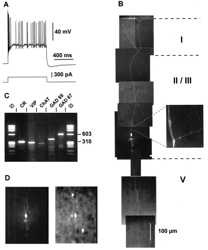
Physiological, morphological, and molecular analyses of a layer II–III IS cell. A, Current-clamp recording obtained in response to a depolarizing current pulse (300 pA). Note the initial burst followed by irregularly discharged action potentials. B, Reconstruction of the cell labeled with biocytin injection revealed a typical bipolar morphology with two narrow vertical dendritic arborizations. The ascending dendrite (top) extended up to layer I where it branched before reaching the cortical surface. The descending dendrites extended to layer V. Inset, Higher magnification of a portion of the proximal ascending dendrite showing the beginning of the axon running horizontally. Note the ascending and descending colaterals displaying varicosities. C, Agarose gel analysis of products of the RT-mPCR designed for the detection of CR, VIP, ChAT, GAD65, and GAD67 mRNAs. The five amplified fragments had the sizes (in bp) predicted by the mRNA sequences of 309 (CR), 287 (VIP), 262 (ChAT), 391 (GAD65), and 600 (GAD67).D, Comparison of the biocytin labeling of the recorded neuron (left panel) with the immunostaining of the slice with an antibody against CR (right panel). Note the immunoreactivity of the biocytin-labeled cell.