
Fig. 2.
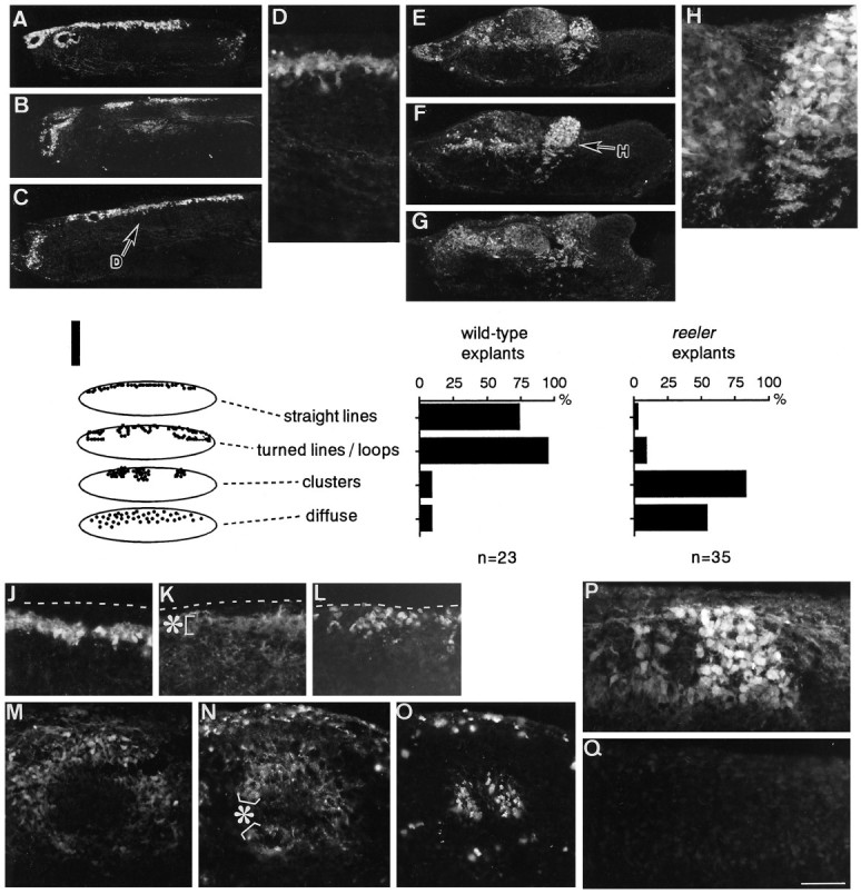
Untreated cerebellar explants derived from normal and reeler mice. Explants fixed at 7 DIV were sectioned across the long axis and stained with anti-calbindin. Sets of photomicrographs show distinct distribution patterns of Purkinje cells between a normal-derived explant (A–D) and areeler-derived explant (E–H).D and H are magnified views of the indicated portion in C and F, respectively. I, Histograms showing the frequencies of occurrence of four “patterns” (vertical axis; also illustrated schematically on the left), which were extracted from photomicrographs of 10–15 separate sections covering the entire explants, in normal and reeler explants. For example, the wild-type case in A–D shows “straight lines” (A–D) and “turned lines” or “loops” (A, B), whereas the reeler case inE–H shows “clusters” (F, H) and a “diffuse” pattern (E, G). One explant often had two or three patterns among the sections, and even one of the sections occasionally showed combined patterns. Therefore, the percentage for one of the patterns is calculated and presented independent of that for the remaining patterns, and the sum of the percentages exceeds 100%. J–Q are magnified views of superficial areas in explants (same magnification), and they show the spatial relationship between Purkinje cells and other layer-like structures identified in vitro. J–O, Sets of photomicrographs showing the relationships between calbindin+ Purkinje cells (J, M), extracellular Reelin/CR-50 antigen (asterisks inK and N), and cells taking up BrdU (L, O) around the “straight line” (J–L) and “loop” (M–O), two PCL-like structures in wild-type explants. The wild-type explants were exposed to BrdU for 1 hr before fixation, and serial sections were stained with antibodies to these markers. In both structures, there was a lamination sequence from a layer or mass of cells taking up BrdU (L, O) to the “straight line” (J) or “loop” (M) formed by Purkinje cells, through an extracellularly CR-50 immunoreactive zone (K, N), which often overlapped the Purkinje cells. Broken lines inJ–L indicate the upper (pial) surface of explants. CR-50 immunoreactivity other than the bands (asterisk) in K andN seems to correspond to granule neurons labeled intracellularly. P, Q, Double staining with anti-calbindin (P) and CR-50 (Q) onreeler explants, in which Purkinje cells were clustered (P) and Reelin was not detected (Q). Scale bar: A–C, E–G, 200 μm;D, H, J–O, 50 μm.