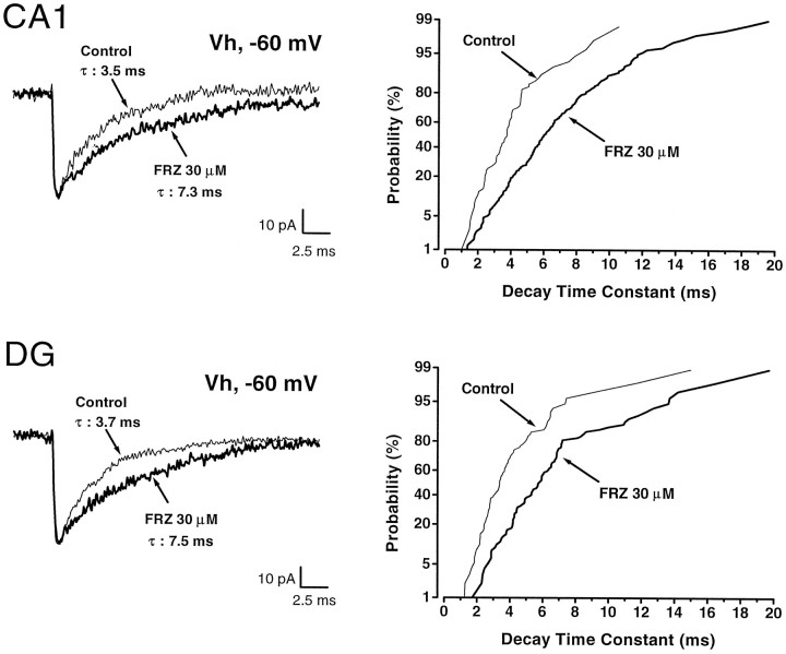
Fig. 2.
Effect of FRZ (30 μm) onCA1 and dentate gyrus (DG) granule cell GABAA receptor-mediated mIPSCs. The left panels represent superimposed averages of 30–35 mIPSCs in control (thin line) and FRZ (thick line) traces. Note that FRZ at concentrations corresponding to “low dose” chronic treatment (30 μm) induced prolongation of the decay time constant from 3.5 to 7.3 msec and from 3.7 to 7.5 msec inCA1 pyramidal cells and DG, respectively. The right panels represent the cumulative probabilities of 92–235 mIPSC decay time constants in the same cells. In both cases, i.e., CA1 and DG, FRZ increased the decay time constants of all mIPSCs. Holding potential was −60 mV.

