
Fig. 8.
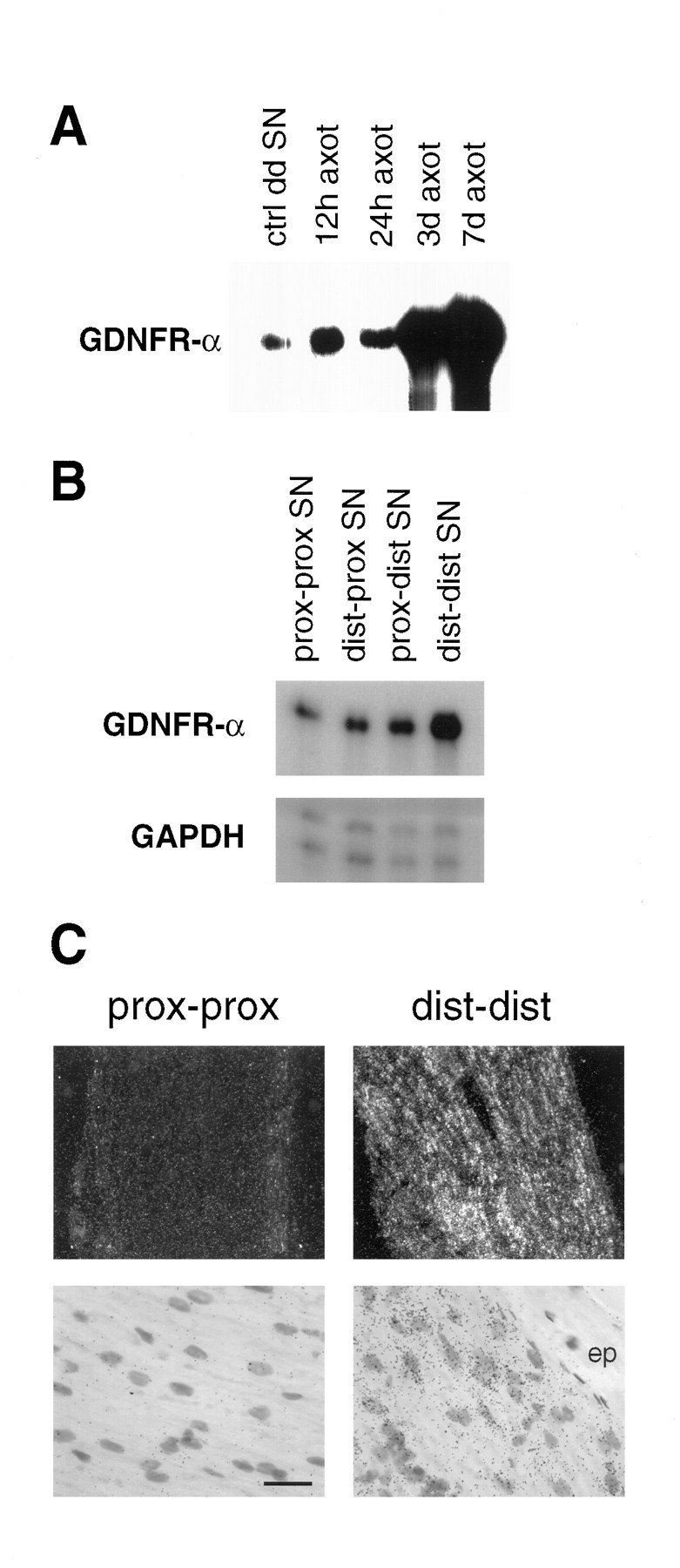
Upregulation of GDNFR-α mRNA after sciatic nerve transection. A, Autoradiogram of an RPA revealing a pronounced increase in GDNFR-α mRNA in the distal segment of transected sciatic nerve 3 and 7 d after surgery.B, A distally increasing gradient of GDNFR-α mRNA is demonstrated in an autoradiogram of an RPA of sciatic nerve samples collected 3 d after nerve transection. C, Photomicrographs of GDNFR-α in situ hybridization of nerve segments from animals sacrificed 3 d after nerve transection. The GDNFR-α mRNA-expressing cells are clearly present in the Schwann cell layer, whereas the epineurium (ep) is devoid of labeling. For analysis, the transected nerve was dissected into four parts: moving laterally from the cord, the proximal segment of the proximal stump (prox-prox), distal segment of the proximal stump (dist-prox), proximal segment of the distal stump (prox-dist), and distal segment of the distal stump (dist-dist). Scale bar (shown inC): dark field, 134 μm; bright field, 18 μm.