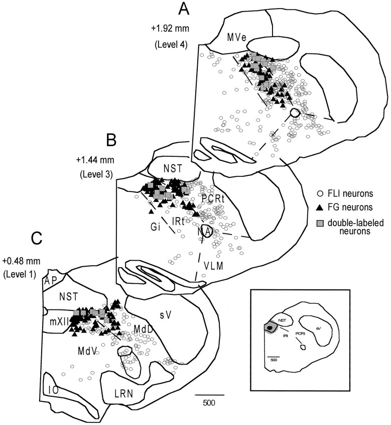
Fig. 6.
Distribution of FLI (open circles), FG-labeled (filled triangles), and double-labeled (shaded squares) neurons throughout the medullary reticular formation for a representative quinine animal. Sections are ordered from rostral (A) to caudal (C).Numbers on the left indicate distance from obex. The inset depicts the location of the FG injection site within the hypoglossal nucleus.

