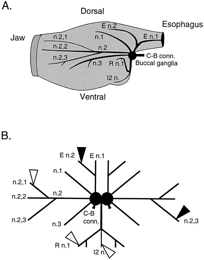
Fig. 1.
The buccal ganglia and its peripheral nerves. A, Schematic representation of the buccal mass and the location of the buccal ganglia and its peripheral nerves. B, Schematic representation of in vitro buccal ganglia preparation showing the position of the recording electrodes (white triangles) on I2 n., n.2,1, and R n.1 and the stimulating electrodes (black triangles) on E n.2 and n.2,3. This schematic illustrates the placement of electrodes that was used in nondesheathed preparations (i.e., the E n.2 that was stimulated was ipsilateral to the nerves from which recordings were made). In desheathed preparations the E n.2 that was contralateral to the ganglion from which recordings were made was stimulated (data not shown). C-B conn., Cerebrobuccal connectives; E n., esophageal nerve; I2 n., nerve to intrinsic buccal muscle 2; n., buccal nerve; R n., radular nerve.

