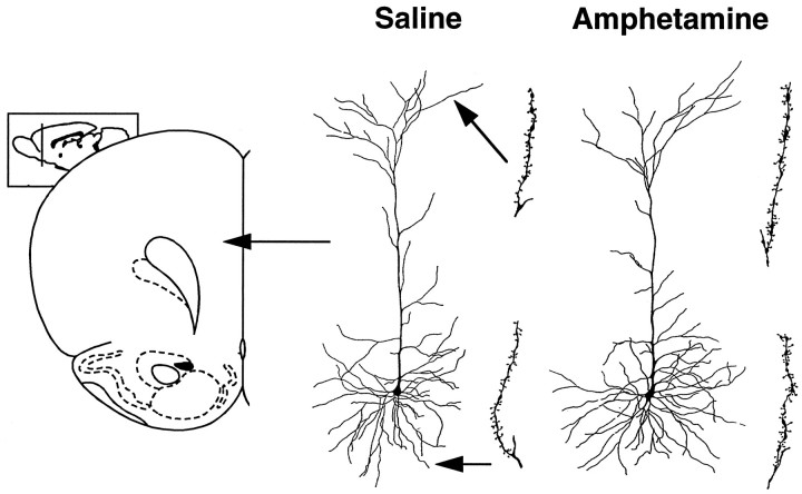
Fig. 4.
Camera lucida drawings of representative layer III pyramidal cells in the prefrontal cortex (area Cg 3) of saline- and amphetamine-pretreated rats. The drawing to the right of each cell represents an apical or basilar dendritic segment used to calculate spine density. Coronal drawing is adapted from Paxinos and Watson (1997).

