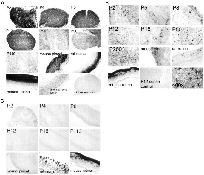
Fig. 1.
Expression of visual pigment mRNA in rodent pineal at different ages. A, Rhodopsin. Rat rhodopsin is used as a probe. All magnifications are at 100× except P2 rat pineal, adult mouse pineal, adult rat retina, and adult mouse retina, which are at 200×. Exposure times for adult rat and mouse retina are 30 min, whereas exposure times for pineals are 2 d. Exposure times for both sense controls are 2 d.B, Blue cone pigment. Mouse blue cone pigment is used as a probe. All magnifications are at 100× except P2 rat pineal, adult mouse pineal, adult rat retina, and adult mouse retina, which are at 200×. The dark band above the photoreceptor layer of the mouse retina in this and all subsequent figures is the retina pigmented epithelium, which is unpigmented in the albino rat retinas tested. The final panel, showing P200, is taken at 400× magnification. C, Red cone pigment. Mouse red/green cone pigment is used as a probe. All magnifications are at 100× except P2 rat pineal, adult mouse pineal, adult rat retina, and adult mouse retina, which are at 200×.

