
Fig. 7.
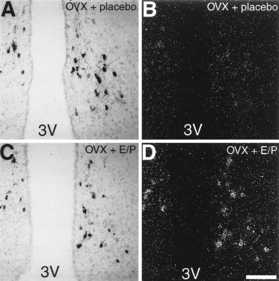
Photomicrographs showing tyrosine hydroxylase-like immunoreactivity (A, C) and AT1A receptor mRNA (B, D) in neurons of the arcuate nucleus of placebo-treated OVX rats (A, B) and estrogen/progesterone-treated OVX rats (C, D). Sections were double-labeled for AT1A receptor mRNA and tyrosine hydroxylase-like immunoreactivity. B andD are dark-field views of A andC, respectively. 3V, Third ventricle. Scale bar, 100 μm (applies to all panels).