
Fig. 1.
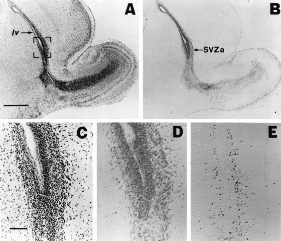
m-Msi-1 expression in P3 forebrain. Serial sagittal sections of the forebrain at P3 with hematoxylin–eosin staining (A, C), immunolocalizations of m-Msi-1 (B, D), and PCNA (E). Anterior is to the right and dorsal is up. C–E, Higher magnifications of the anterior corner of the lateral ventricle (SVZa) shown in the bracketed portion ofA. m-Msi-1 immunostaining was observed in PCNA-positive proliferating cells in SVZa. Note that m-Msi-1 expression was also detected in the posterior SVZ in addition to the migratory route of neuronal precursor cells from SVZa into the olfactory bulb described previously (Luskin, 1993). Scale bars: A, B, 500 μm;C–E, 50 μm. SVZa, Anterior region of the SVZ; lv, lateral ventricle.