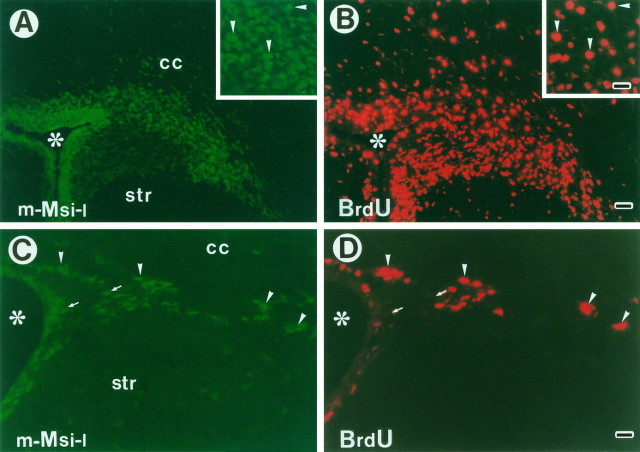
Fig. 2.
m-Msi-1 expression in proliferating cells residing in the postnatal and adult SVZ. To label the entire constitutively proliferating population surrounding the lateral ventricles, P7 and adult mice received 3 and 12.5 hr of BrdU injections, respectively. Double-immunofluorescent labeling of coronal sections through the SVZ surrounding the lateral ventricle in P7 forebrain (A, B) and adult forebrain (C, D) with antibodies to m-Msi-1 (A, C; FITC) and BrdU (B, D; Cy3). Lateral is to the right and dorsal is up. Insets inA and B, Higher magnifications of the dorsolateral corner of the P7 SVZ. Many dividing, small, densely packed cells that are brightly immunostained with both m-Msi-1 and BrdU are observed in the postnatal developing SVZ (arrowheads ininsets in A and B). Similarly, a considerable number of m-Msi-1- and BrdU-positive dividing cells are also present in the adult subependyma (arrowheads in C and D), although it is obvious that there is a subpopulation of m-Msi-1-positive but BrdU-negative cells (arrows inC and D). Scale bars: A, B, 36 μm; insets, C and D, 18 μm. Asterisks, Lateral ventricle; cc, corpus callosum; str, striatum.

