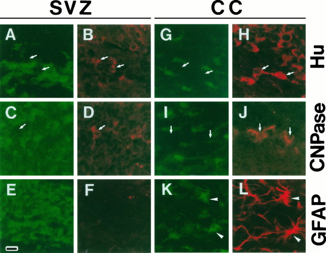
Fig. 3.
Cell type of m-Msi-1-positive cells in the developing SVZ and the corpus callosum. Double-labeling of coronal sections just dorsolateral to the region of the P7 SVZ (A–F) and the P7 corpus callosum (G–L) with anti-m-Msi-1 antibody (A, C, E, G, I, and K; FITC), anti-Hu (B, H; Cy3), anti-CNPase (D, J; Cy3), and anti-GFAP (F, L; Cy3). The bar in Erepresents 18 μm. Antibodies to CNPase and Hu label populations that are distinct from m-Msi-1-positive cells in both the SVZ and the corpus callosum. The arrows point to the Hu-positive but m-Msi-1-negative cells (A, B, G, H), and CNPase-positive but m-Msi-1-negative cells (C, D, I, J). Although GFAP immunoreactivity is not detected in the SVZ, a few m-Msi-1- and GFAP-positive cells that have multiple processes or short branched processes are now present in the developing corpus callosum (arrowheads in K andL) among the more numerous m-Msi-1-positive cells.

