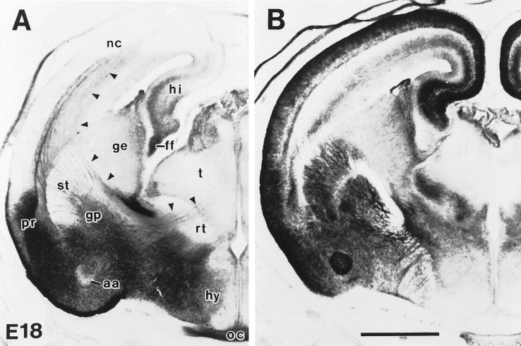
Fig. 3.
GLT-1 (A) and EAAC1 (B) immunoreactivity in rat forebrain at E18. GLT-1 was expressed in the amygdala, perirhinal cortex, hippocampus, fimbria, and optic nerve; however, sparse immunoreactivity was seen in embryonic neocortex and striatum. GLT-1 immunoreactivity was also seen along white matter pathways interconnecting neocortex, basal ganglia, and thalamus (arrowheads). In contrast, strong EAAC1 immunoreactivity was seen in neocortex, striatum, hippocampus, and reticular thalamic nucleus but not in the fimbria and optic nerve.aa, Anterior cortical amygdaloid nucleus;ff, fimbria fornix; ge, ganglionic eminence; gp, globus pallidus; hi, hippocampus; hy, hypothalamus; nc, neocortex; oc, optic chiasm; pr, perirhinal cortex; rt, reticular thalamic nucleus;st, striatum; t, thalamus. Scale bar, 1 mm.

