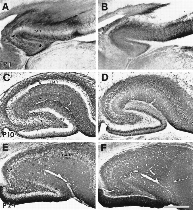
Fig. 6.

Immunoreactivity for GLT-1 (A, C, E) and EAAC1 (B, D, F) in parasagittal sections of hippocampus at P1 (A, B), P10 (C, D), and P24 (E, F). GLT-1 and EAAC1 immunoreactivities were seen throughout development and into adulthood. Postnatally, GLT-1 was localized in astrocytes. EAAC1 was seen in neurons and was more enriched in pyramidal and granule cells at P10 (D). CA1 and CA3, Cornu Ammon’s subfields 1 and 3; dg, dentate gyrus;H, hilus; o, stratum oriens;p, stratum pyramidale; r, stratum radiatum; lm, stratum lacuosum-moleculare;m, molecular layer; g, granular layer. Scale bar, 500 μm.
