
Fig. 8.
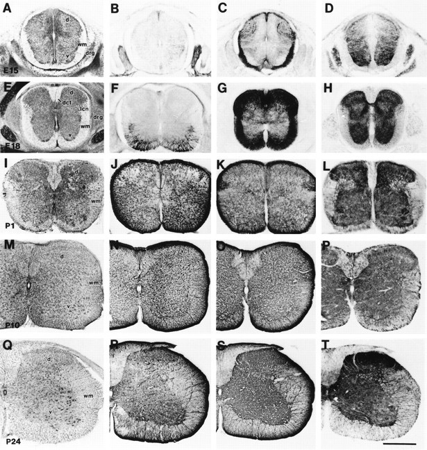
Cresyl violet staining (A, E, I, M, Q) and immunoreactivity for GFAP (B, F, J, N, R), GLT-1 (C, G, K, O, S), and EAAC1 (D, H, L, P, T) in cervical spinal cord at E15 (A–D), E18 (E–H), P1 (I–L), P10 (M–P), and P24 (Q–T). GFAP was expressed at E18 in the medioventral white matter (F). Intense GLT-1 immunoreactivity was seen in white matter and dorsal horn at E15 (C) and E18 (G), although GLT-1 was more expressed in astrocytes of gray matter after P10 (O, S). EAAC1 was localized in gray matter throughout embryonic to postnatal periods. d, Dorsal horn;v, ventral horn; wm, white matter;drg, dorsal root ganglion; dct, dorsal corticospinal tract; lcn, lateral cervical nucleus. Scale bar, 500 μm.