
Fig. 5.
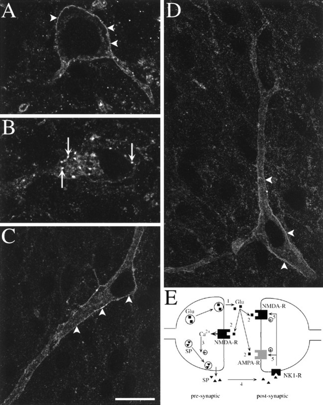
Fluorescent confocal images of NK1R-immunoreactive neurons in slices electrically stimulated. Stimulation consisted of three trains of 1 sec at 100 Hz separated by 10 sec, delivered to one of the dorsal roots. Arrowheads indicate surface labeling, and arrows indicate internalized NK1R.A–C, Single optical sections. A, Lamina I neuron in the contralateral side showing surface immunoreactivity.B, Lamina I neuron in the stimulated side showing extensive NK1R internalization. C, Lamina III neuron in the contralateral side. D, Lamina III neuron in the stimulated side with a dendrite projecting to lamina I (at thetop); this is a computer-generated juxtaposition of two confocal images of four (top) or five (bottom) optical sections. Scale bar: A, B, 10 μm; C, D, 20 μm. E, Model of the synergistic action of SP and glutamate at a dorsal horn synapse. 1, Noxious stimuli produce presynaptic depolarization and glutamate release. 2, Presynaptic NMDA receptors are activated by glutamate and depolarization, letting Ca2+ inside the presynaptic button.3, Increases in presynaptic calcium concentration trigger SP release. 4, SP binds to NK1Rs in surrounding neurons starting the internalization process. 5, Activated NK1Rs increase postsynaptic responses to glutamate.