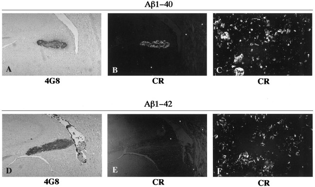
Fig. 1.
Photomicrographs of rat brain sections showing aggregates formed after intracerebral injection of freshly solubilized Aβ1–40 (A, B) and Aβ1–42 (D, E) at 3 weeks (A, B) and 1 d (D, E) postinjection survival times. The sections were probed by immunohistochemistry with 4G8 (A, D) and by Congo red staining (CR) (B, E). The preparations of Aβ1–40 (C) and Aβ1–42 (F) were preincubated in vitro to form fibrils, followed by staining with Congo red. Photomicrographs (B, C, E, F) were taken under illumination with polarized light.

