
Fig. 2.
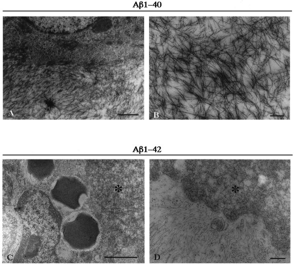
Electron micrographs of rat brain injected with freshly solubilized Aβ1–40 (A, B) and Aβ1–42 (C, D) at 1 week postinjection survival time. Injected Aβ1–40 consists of 5–10 nm fibrils, and injected Aβ1–42 consists of nonfibrillar amorphous material (indicated as *). Note that Aβ1–42 amorphous aggregates are intimately intermingled with macrophages (C) and astrocytes (C, D). Scale bars: A, 1 μm; B, 250 nm; C, 1 μm; D, 250 nm.