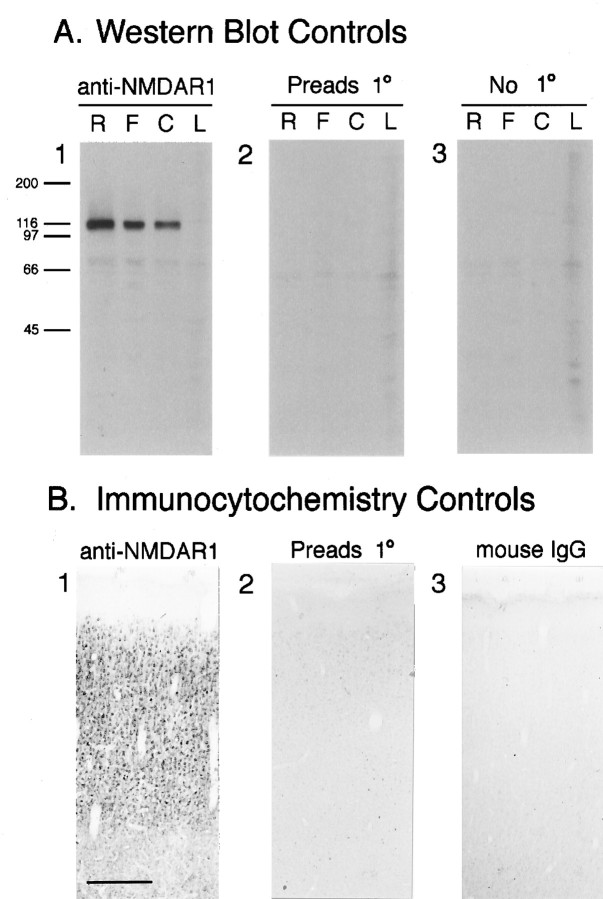
Fig. 1.
A, Western blots show that the monoclonal antibody recognizes a band of the same molecular weight as NMDAR1. A1, Staining of rat (R), ferret (F), and cat (C) synaptic plasma membranes and ferret liver membranes (L) with the primary anti-NMDAR1 antibody yields a prominent band at Mr ∼117 kDa. This band is no longer stained when the primary antibody is preadsorbed to its fusion protein immunogen (A2), or omitted (A3). B, Controls for the specificity of NMDAR1 antibody immunostaining on tissue sections. Cellular staining is intense in sections that have been incubated in primary antibody (B1); however, preadsorbtion of the primary antibody with the fusion protein that it was raised against (B2) or substitution of the primary antibody with generic IgG protein from the same species as the primary (mouse IgG) (B3) protein results in the near-absence of cellular staining. Scale bar (B1–3): 300 μm.

