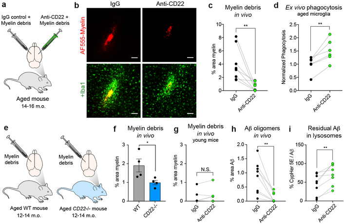
Figure 3. CD22 inhibition restores microglial phagocytosis in vivo.
a, Myelin debris labeled with a fluorescent dye (AF555) was stereotactically co-injected with anti-CD22 or IgG into the cortex on opposite hemispheres of the same aged (14-16 m.o.) mouse.
b, Representative images of AF555-labed myelin (red, top row) overlaid with the myeloid marker Iba1 (green, bottom row) at the injection sites of IgG (left) or anti-CD22 (right) treated hemispheres of the same aged brain. Scale bar = 100 microns.
c, Clearance of myelin debris in IgG (black) or anti-CD22 (green) treated hemispheres of aged mice assessed 48 hours post-injection (n=8, **P<0.005, paired two-sided t-test).
d, Flow cytometry quantification of ex vivo phagocytosis of pH-sensitive beads by aged microglia pretreated with IgG or anti-CD22 (n=6, **P<0.005, paired two-sided t-test).
e, Labeled myelin debris was stereotactically injected into the cortices of aged (12–14 m.o.) WT or CD22−/− mice.
f, Clearance of myelin debris in cortices of aged WT (black) vs CD22−/− (green) mice assessed 48 hours post-injection (n=4, *P<0.05, two-sided t-test, mean +/− s.e.m.).
g, Clearance of myelin debris in IgG (black) or anti-CD22 (green) treated hemispheres of young mice assessed 48 hours post-injection (n=4, N.S. not significant, paired two-sided t-test).
h, Clearance of Aβ oligomers in IgG (black) or anti-CD22 (green) treated hemispheres of aged mice assessed 48 hours post-injection (n=8, **P<0.005, paired two-sided t-test).
i, Percent area of residual Aβ oligomers that were CypHer5E+, indicating localization to acidified lysosomes (n=8, **P<0.005, paired two-sided t-test).
All data were replicated in at least 2 independent experiments.

