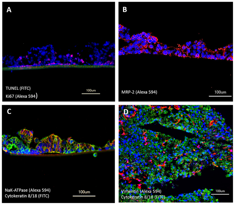
Fig. 2.
Performance of liver–intestine co-cultures in MOCs over 14 days. Staining for (A) TUNEL/Ki67 showing apoptosis (green) and viability (red) in small intestinal epithelial tissue. (B) MRP-2 (red) expression in small intestinal epithelial tissues. (C) Transporter NaK-ATPase (red) and epithelial cells cytokeratin 8/18 (green) in small intestinal epithelial tissues. (D) Equal distribution of HHSteC, stained with vimentin (red), within hepatocytes, stained with cytokeratin 8/18 (green). (A–D) Nuclei were stained with DAPI (blue).

