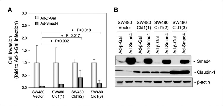
Figure 4.
The effect of overexpression of claudin-1 on Smad4-mediated tumor-suppressive function in SW480 cells. A, cells grown on six-well plate to 50% to 60% confluency were infected with β-galactosidase or Smad4 adenovirus at a multiplicity of infection of 20 as described in Materials and Methods. Forty-eight hours after infection, cells were plated in Matrigel-coated Transwells (100,000 cells per Transwell) and allowed to grow for 3 days. Invasive cell numbers were scored as described in Materials and Methods, and the results are presented as fold change to β-galactosidase adenoviral infection. Columns, mean fold change; bars, SD. *, P < 0.05 compared with control. The P value was determined using Student’s t test. B, cells were infected with β-galactosidase or Smad4 adenovirus at a multiplicity of infection of 20. Seventy-two hours after infection, cells were lysed, and the lysates were subjected to immunoblotting for Smad4, claudin-1, and β-actin.

