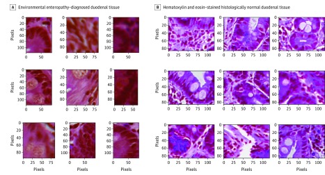
Figure 2. High Activation Areas .
A, Hematoxylin-eosin–stained duodenal tissues with diagnosed environmental enteropathy (original magnification ×100). B, Hematoxylin-eosin–stained histologically normal duodenal tissue (original magnification ×100). These images were the areas of high activation identified by the model; we observed secretory cells, specifically Paneth cells and goblet cells, in the mucosa. Our classification model identified these secretory cells to be of high importance for distinguishing biopsies with no disease from biopsies of environmental enteropathy and celiac disease.

