Fig. 4.
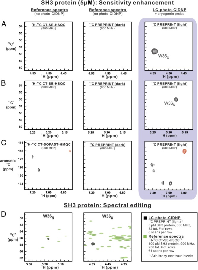
The 2D 13C-PREPRINT analysis of the SH3 protein (5 μM), highlighting the benefits of laser-enhanced NMR. (A) Upfield and (B) downfield Hα-Cα regions of 2D heteronuclear correlation spectra of the SH3 protein. The 2D 13C PREPRINT spectrum (16 complex points in indirect dimension, eight scans per row) under light (i.e., laser-on) conditions is shown along with reference spectra (1H-13C CT-SE-HSQC and dark 13C PREPRINT, i.e., under laser-off conditions) collected with identical number of scans per row, recycle delays, sweep widths, and number of points in both dimensions. To highlight the power of photo-CIDNP, LC-photo-CIDNP and 1H-13C CT-SE-HSQC data were collected at 600 and 900 MHz, respectively. (C) The 2D spectra analogous to A and B, except that the focus is on the 1H-13C aromatic region. In this case, 1H-13C CT-SOFAST-HMQC was employed as a reference experiment. To achieve an identical total experiment time, the 1H-13C CT-SOFAST-HMQC experiment included 36 scans per row. (D) Overlay of 13C PREPRINT 2D spectrum of the SH3 protein (5 μM; black) and the 2D 1H-13C CT-SE-HSQC reference spectrum (100 μM; green). The overlay highlights the dramatic spectral-editing capabilities of 13C PREPRINT, enabling the rapid selective detection of photo-CIDNP-active resonances of complex macromolecules.

