Fig. 1.
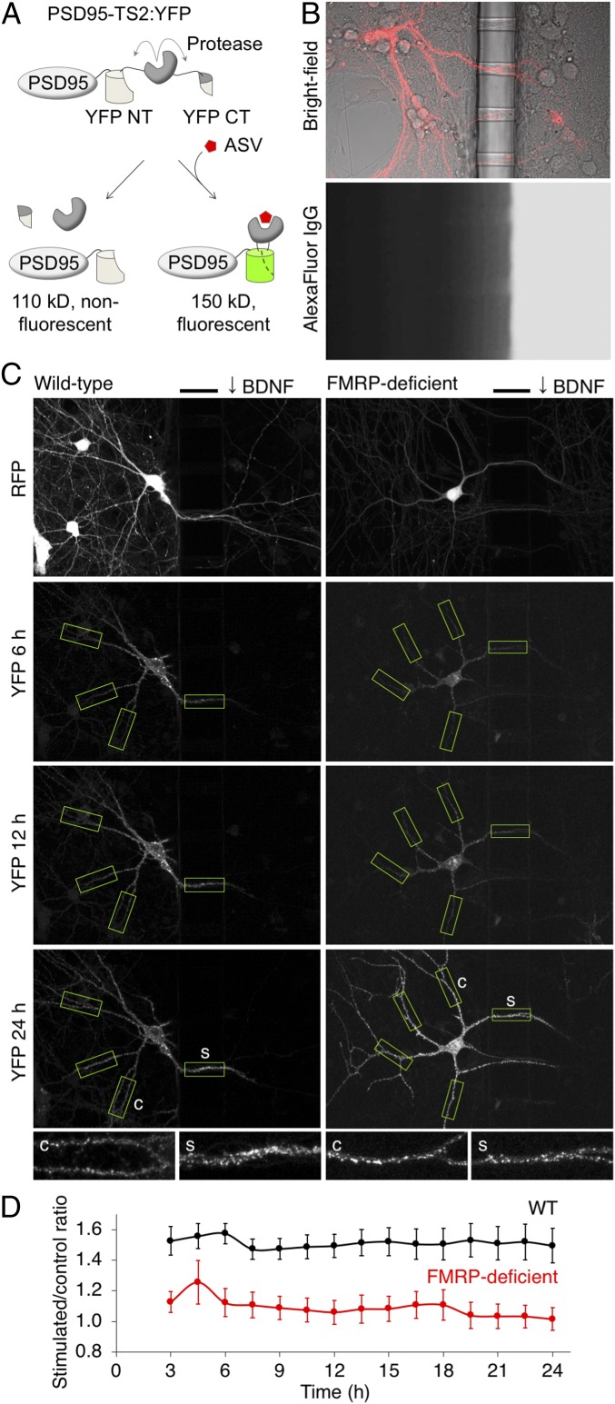
Local expression of new PSD95 in BDNF-stimulated dendritic regions is absent in FMRP-deficient neurons. (A) Diagram of the PSD95-TS2:YFP reporter. After asunaprevir is added, YFP fluorescence is preserved on new PSD95 copies. CT, C terminus; NT, N terminus. (B) Compartmentalized culture system. (B, Top) Superimposed fluorescence and bright-field images showing a transfected neuron extending dendrites through tunnels in the barrier. (B, Bottom) Dye added to one side for 24 h demonstrates separation between compartments and dye diffusion down the tunnels. Barrier width is 50 μm. (C) New PSD95 in WT or FMRP-deficient neurons after local BDNF stimulation. The bars mark the location of the 50-μm barrier. BDNF was added to the right chamber. Boxes indicate regions identified and analyzed by the automated algorithm. Stimulated segments were defined as the 50 μm in the tunnel, and then control segments were defined in the unstimulated compartment at an equidistant path length from the cell body. (C, Insets) Stimulated (s) and control (c) segments. (D) Stimulated/control intensity ratios in FMRP-deficient neurons were significantly lower than in WT neurons at all times (P = 0.03 by mixed-effect repeated-measures ANOVA; n = 31 WT and 24 FMRP-deficient neurons). Error bars represent SEM.

