
Figure 6.
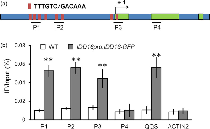
IDD16 directly associates with the SPCH promoter. (a) SPCH locus comprising the 2‐kb promoter region and transcribed region. Black lines denote fragments amplified in ChIP‐qPCR (b). Red bar represents the core binding sites (TTTGTC or GACAAA) of IDD proteins. (b) qPCR of fragments (as in a) from ChIP of IDD16pro:IDD16‐ GFP seedlings and WT with anti‐GFP antibody. Negative control P4 for ChIP‐qPCR are located in the second exon. Values are means ± SE from three biological replicates. Asterisks indicate statistical significance based on Student's t test; ** P < 0.01.