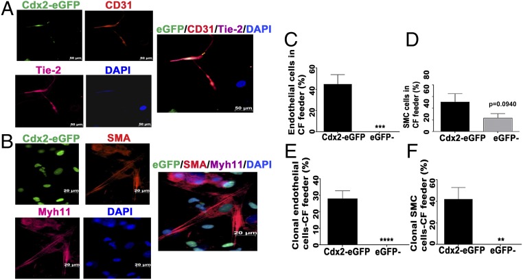
Fig. 3.
Vascular differentiation of Cdx2-eGFP cells in vitro. Cdx2-eGFP cells differentiated into endothelial (CD31/Tie-2) cells (A) and smooth muscle (αSMA/myh11) cells (B) in vitro (CD31 and SMA, red; Tie-2 and Myh11, pink; DAPI, blue). (Scale bar: 20 μm.) Quantification of Cdx2-eGFP cells derived from endothelial cells (C) and smooth muscle (D) cells in vitro in CF feeders. (E and F) Quantification of clonal differentiation of Cdx2-eGFP cells into vascular lineages. However, eGFP− cells did not display clonal vascular commitment compared with the Cdx2-eGFP cell population. Data are represented as mean ± SEM (n = 3 independent experiments). **P = 0.0065, ***P = 0.005, ****P < 0.0001.

