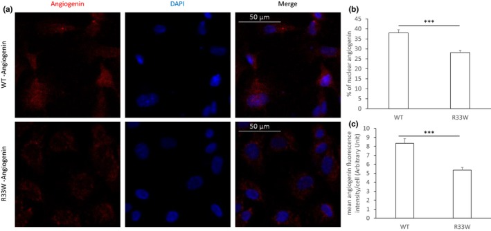
Figure 5.

Nuclear translocation of wild‐type (WT) and mutant angiogenin (ANG) in human umbilical vein endothelial cells (HUVEC) cells. HUVEC cells were incubated with 1 μg/ml of WT ANG or mutant ANG for 30 min and subsequently immunostained for ANG. Nuclei were stained with 4′,6‐diamidino‐2‐phenylindole (DAPI). Images show the representative result of four experiments. (a) WT ANG accumulates both in the cytoplasm and nucleus of the HUVEC cells, whereas mutant (R33W) ANG accumulated predominantly in the cytoplasm of the cells. Individual channels as well as merged images are shown. (b) Nuclear translocation was quantified by measuring mean signal intensity levels of ANG staining in nucleus and cytoplasm of cells for at least three fields of views. Quantification of nuclear translocation of WT and mutant (R33W) ANG showed a significant decrease in nuclear staining in the R33W‐treated samples (two‐tailed Student's t‐test, p < 0.0001). (c) Mean cellular signal intensity levels of ANG was determined for at least three fields of views, which showed a significant difference between the intensity of staining of the WT and mutant protein (two‐tailed Student's t‐test, p < 0.0001), suggesting a difference in the cellular internalization of the protein
