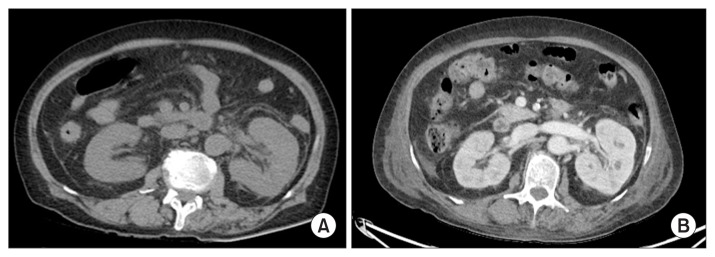
Figure 1. Abdominal computed tomography (CT) findings.
(A) At admission, a non-enhanced CT showed swelling of the left kidney with perirenal fat stranding and renal fascial thickening, suggestive of acute pyelonephritis. (B) Follow-up enhanced CT on day 6 showed multiple small abscesses in the left kidney.

