
Figure 4.
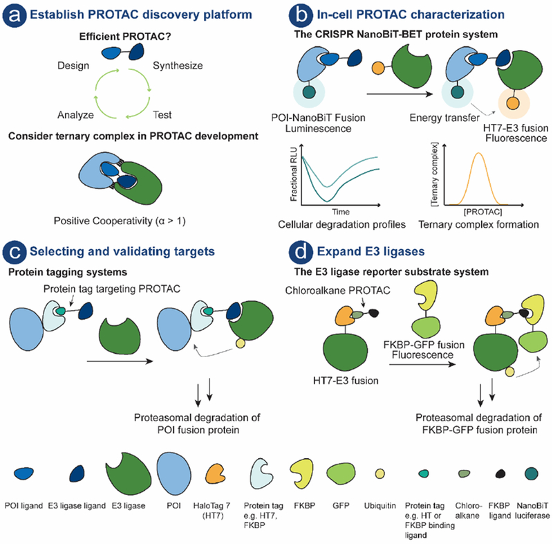
a) PROTAC compound; POI ligand (royal blue), E3 ligand (dark blue) connected by a linker (black). PROTAC platform with iterative design, synthesis, test and analyze cycle. b) Schematic illustration of the CRISPR NanoBiT-BET protein system [86]. This system enables PROTAC characterization in live-cells using endogenous protein levels. c) Protein tagging systems can be exploited for target selection and/or validation. The POI is fused to e.g. HT7 [61] or FKBP[87] and PROTAC mediated ternary complex formation results in polyubiquitination of the POI followed by proteasomal degradation. d) System for assessing E3 utility in the PROTAC technology [88]. Schematic illustration of the E3 reporter substrate system. E3 is fused to HT7, chloroalkane PROTAC covalently binds to HT7, and the recruitment of the reporter substrate (FKBP-GFP fusion protein) results in ternary complex, polyubiquitination and subsequent proteasomal degradation of the reporter substrate.