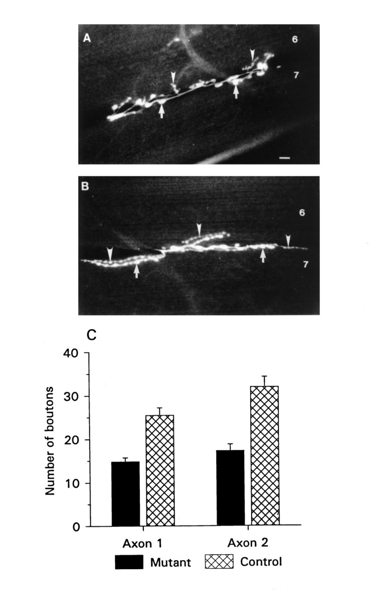
Fig. 1.

Aberrant neuromuscular morphology in Fas II mutants. A, B, Fluorescence micrographs of NMJs of ventral longitudinal muscles 6 and 7 in mutant (A) and control (B) animals. Arrows point to varicosities of axon 1, and arrowheads point to varicosities of axon 2. Scale bar (shown in A), 10 μm. C, Summary of varicosity counts obtained from 18 mutant and 13 control NMJs from abdominal segment 4. The error bars represent the SEM in this and subsequent figures. Mutant and control animals are from the e76 and e93 P-element excision lines described in Grenningloh et al. (1991).
