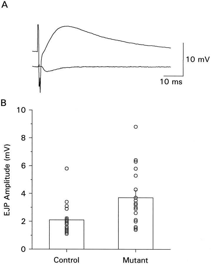
Fig. 7.
EJPs recorded with focal calcium application. A micropipette containing 2 mm calcium was placed over several regions of the nerve terminal on each muscle fiber and covered areas of both varicosity types. The segmental nerve was stimulated at a voltage to recruit both axons, and EJPs were recorded with an intracellular electrode. The bathing solution contained 0 calcium. A, Example of raw trace showing evoked EJP (top trace) and synaptic event recorded through the focal pipette (bottom trace). B, The bar graph shows the mean EJP amplitude of data collected from mutant (n = 18) and control (n = 19) sites. The symbolsrepresent the results obtained from individual recording sites.

