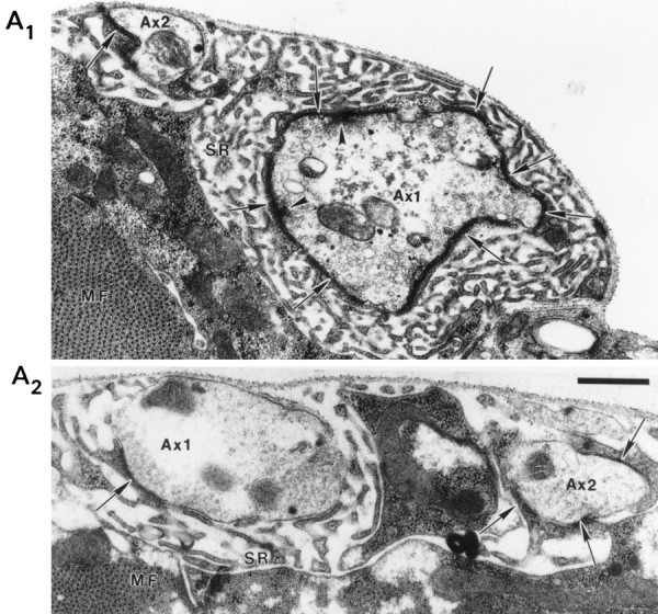
Fig. 8.

Nerve terminal ultrastructural of a Fas II mutant. Electron micrographs of mutant (A1) and control (A2) larval NMJ from abdominal segment 4 showing densely staining synapses (arrows), presynaptic dense bodies (arrowheads), subsynaptic reticulum (SR), and muscle fibers (MF). Axons 1 and 2 are labeled Ax1 andAx2, respectively. The scale bar is 0.5 μm and applies to both A1 andA2.
