
Fig. 2.
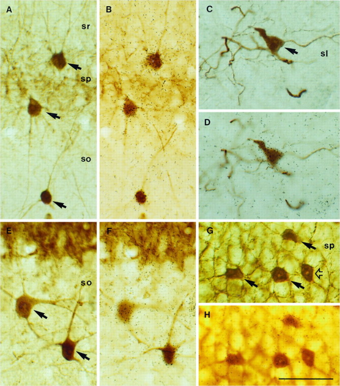
Expression of NGF and NT3 mRNAs in hippocampal interneurons identified with calcium-binding protein immunocytochemistry. Pairs of photomicrographs show the same fields focused at either the plane of the tissue sections or the overlying autoradiographic emulsion. Double-labeled neurons are indicated byarrows. A, B, Several PARV-positive neurons express NGF mRNA in the CA1 region. C, D, One CALR-immunolabeled neuron in the stratum lucidum (sl) of CA3 is overlaid by autoradiographic silver grains after NGF hybridization.E, F, Two CALB-positive neurons in stratum oriens (so) of CA3 show positive hybridization for NGF. G, H, Several PARV-immunopositive neurons in the pyramidal layer (sp) of CA3 express NT3 mRNA; open arrow points to a PARV/NT3-negative interneuron. Scale bar, 50 μm.