
Fig. 5.
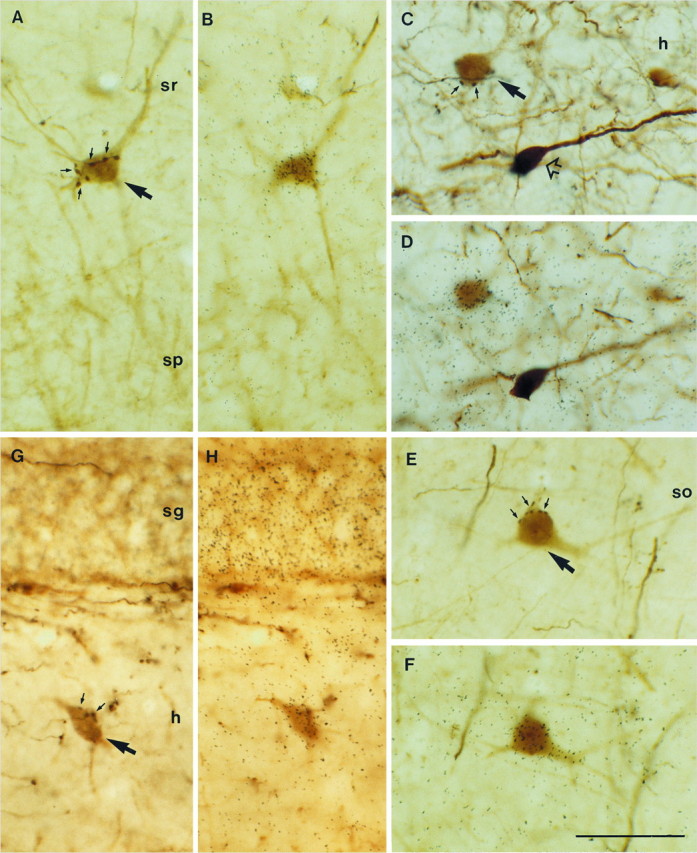
Expression of NGF and NT3 mRNAs in hippocampal interneurons receiving GABAergic septohippocampal input after PHAL injections in the septum. Triple-labeled neurons are indicated bylarge arrows. A, B, A PARV-positive neuron in stratum radiatum (sr) of the CA1 region is contacted by several PHAL-labeled, GABAergic septohippocampal boutons forming a pericellular array (small arrows), and it expresses NGF mRNA. C, D, Expression of transcripts encoding for NGF in a CALR-positive hilar neuron (h) receiving input from GABAergic septohippocampal axons (small arrows). A CALR-positive neuron not innervated by GABAergic fibers is lacking NGF mRNA (open arrow). E, F, A triple-labeled, CALB-positive neuron in stratum oriens (so) of CA3 expresses NGF mRNA. G, H, Expression of NT3 mRNA in a PARV-positive interneuron in the hilar region of the dentate gyrus is surrounded by PHAL-labeled boutons (small arrows) in a basket-like fashion. Notice the dense NT3 hybridization in the granule cell layer (sg). sp, Stratum pyramidale. Scale bar, 50 μm.