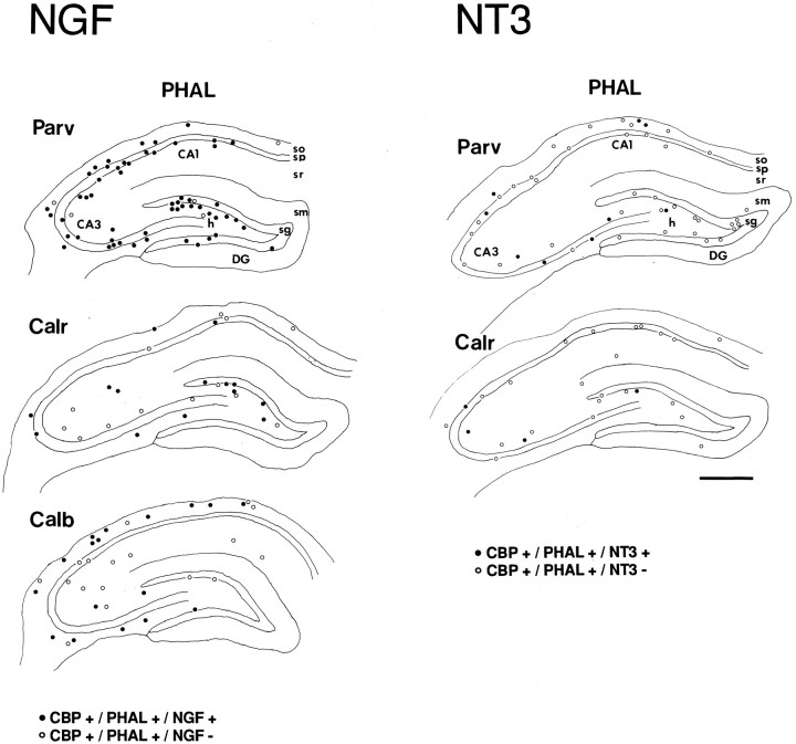
Fig. 6.
Camera lucida drawings showing the distribution of triple-labeled cells (circles) innervated by PHAL-labeled fibers that express either NGF (left) or NT3 (right), and one of the three calcium-binding proteins (CBP). The distribution of neurons showing calcium-binding protein immunostaining and PHAL-labeling but not expression of neurotrophins is indicated by open circles. Plotsrepresent the distribution of cells within one tissue section, except for the CALB-immunoreacted section (two sections). Abbreviations as in Figure 4. Scale bar, 500 μm.

