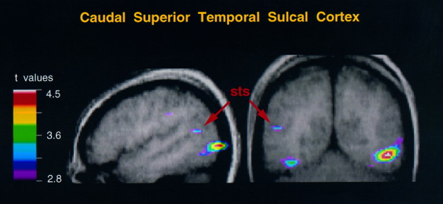
Fig. 1.
Merged PET–MRI sections at x = −48 (sagittal section) and y = −61 (coronal section) to illustrate the activity within the upper bank of the left caudal superior temporal sulcus, in the hand action minus random motion condition. Note that the activity extends into the posterior temporo-occipital region. sts, Superior temporal sulcus. In the coronal section, the subject’s left is on the left side.

