
Fig. 1.
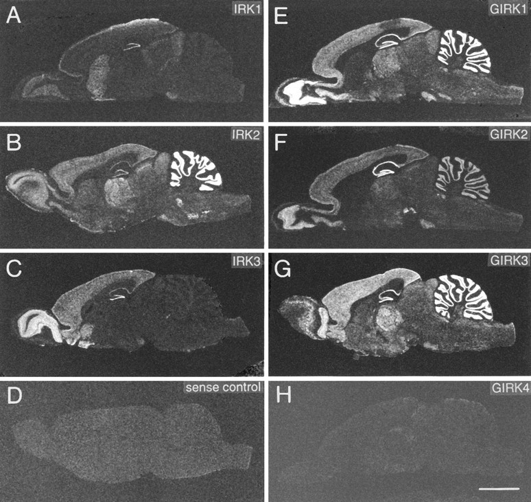
X-ray film images of sagittal rat brain sections show distribution of mRNAs as detected by in situhybridization with oligonucleotide probes specific for IRK1 (A), IRK2 (B), IRK3 (C), GIRK1 (E), GIRK2 (F), GIRK3 (G), and GIRK4 (H). D, Control section hybridized with sense probe. Scale bar (shown in H): 5 mm. Exposure times are 8–21 d.