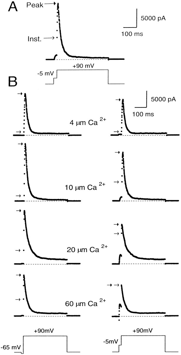
Fig. 2.

Peak and instantaneous currents in BKi cells. In A, 10 μm[Ca2+]i was introduced into the cell. First the cell was stepped from a holding potential of −60 to −5 mV for 20 msec and then stepped directly to +90 mV for 400 msec. After the step to 90 mV, there was a large, ohmic increase in current (the instantaneous current), which was followed by some additional transient activation of current. In B, currents were recorded using pipettes containing 4, 10, 20, or 60 μm free [Ca2+]i. The cells were stepped from the holding potential (−60 mV for cells with 4 and 10 μm[Ca2+]i; −80 mV for cells with 20 and 60 μm[Ca2+]i) to either −65 (left traces) or to −5 mV (righttraces) for 20 msec and then stepped to +90 mV. The instantaneous current at +90 mV became larger at the more positive conditioning steps and increased further as the [Ca2+]i was increased. In all cases, cells were bathed in an external saline containing 0 [Ca2+]o.Arrows on each trace denote the instantaneous (openarrows) and peak (closed arrows) current. Sampling period, 500 μsec.
