Fig. 7.
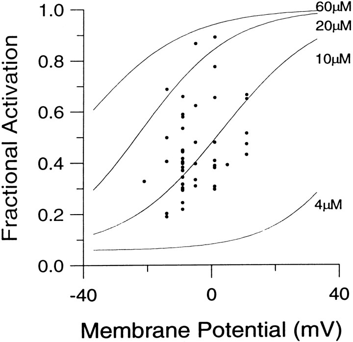
The fractional activation of BKi current elicited by large Ca2+ influx steps corresponds to an average submembrane [Ca2+]i of 10–20 μm. Ca2+ influx in perforated patch-clamped cells was elicited by loading steps between −20 and +11 mV, which were followed by steps to +81 mV. In each cell, the duration of the Ca2+ loading step was increased progressively by using the protocol shown in Figure 5. The fractional activation of BK current for a given Ca2+ influx potential was measured by calculating the ratio of the instantaneous to the peak current for the trace showing the maximal peak BK current. In some cases, traces for which the Ca2+ loading-step durations were longer than the duration that elicited the maximal peak BK current were used. The fractional activation data obtained at various Ca2+ influx potentials from 34 cells are shown here. Solid lines are the fractional activation calibration curves from Figure 4.

