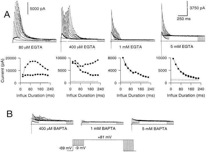
Fig. 8.
EGTA eliminates the slowly developing increase in peak current but not the instantaneous current, whereas BAPTA eliminates all BK current. A, EGTA at concentrations ranging from 80 μm to 5 mm was introduced into cells, and BK current was elicited with Ca2+ influx. Cells were held at −69 mV, stepped to −9 mV to produce Ca2+ influx, and then stepped to +81 mV. The duration of the step to −9 mV was increased from 0 to 220 msec in increments of 20 msec. Shown below are the peak (circles) and the instantaneous (squares) currents plotted against the duration of the Ca2+ influx step. The first voltage step was ignored in this plot, because it does not result in any Ca2+ influx. The dotted linein each case indicates the amplitude of the steady-state Ca2+-independent, voltage-dependent current at +81 mV. As the EGTA concentration is increased, the slowly developing rise in peak current is eliminated. B, BAPTA at concentrations of 400 μm and 1 and 5 mm was introduced into each of three chromaffin cells. BAPTA even at 400 μm is quite effective in eliminating BK current. Note the different y-axis scales in A.

