
Fig. 2.
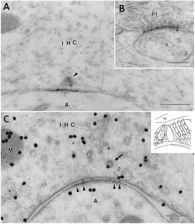
Immunoreactivity for GluR2/3 at an inner hair cell (IHC) synapse in the organ of Corti (A, C) and a parallel fiber (Pf) to Purkinje cell synapse in the cerebellum (B). The 1.4 nm gold particles were made visible by silver enhancement. The section in A is not at the center of the synapse because the synaptic body (arrowhead) is cut near its periphery. C, Double immunolabeling. After demonstration of GluR2/3 by silver intensification (small particles,arrowheads), the sections were immunolabeled for glutamate (30 nm gold particles). Some of the large particles appear to be associated with vesicles (arrow) and with mitochondria (M). Inset shows a diagram of the organ of Corti.Frame indicates area represented in this and subsequent illustrations. Asterisk, Inner hair cell contacted by afferent dendrites; 1–3, the three rows of outer hair cells. s, Purkinje cell spine; A, afferent dendrite; TM, tectorial membrane. A, B, Fixative No. 1 (see Materials and Methods). C, Fixative No. 2. Freeze substitution. Scale bars: 0.5 μm in A, 0.2 μm in B and C.