
Fig. 5.
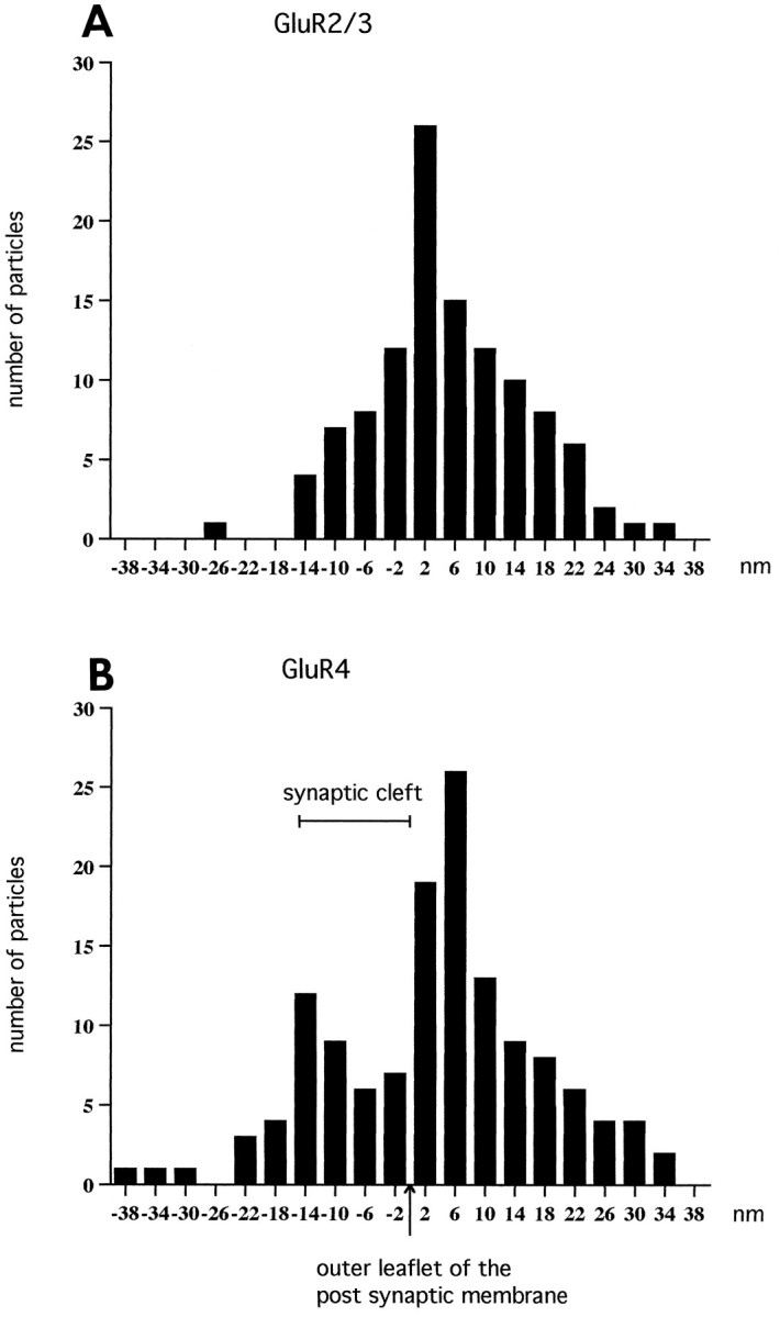
Histograms showing the radial distribution of gold particles representing GluR2/3 (A) and GluR4 (B) at inner hair cell synapses. The distances between the centers of the 15 nm gold particles and the outer leaflet of the postsynaptic membrane were grouped into bins 4 nm wide. The values along the abscissa indicate bin centers. Minus signs indicate direction of the presynaptic element. The data were pooled from 25 synapses (A) and 23 synapses (B). The particles signaling GluR2/3 (A) showed essentially a normal distribution with an average of 4.9 nm (SEM 1.0). The histogram of GluR4 distribution was broader than that of GluR2/3 and displayed two peaks. The extent of the synaptic cleft is indicated.