
Fig. 3.
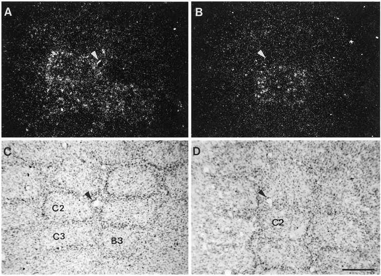
One-to-one relationship between the stimulated whiskers and the corresponding contralateral, stimulated barrels.A and B are dark-field photomicrographs showing BDNF mRNA hybridization signal in tangential sections through stimulated barrel cortices of 6 hr stimulated animals. In A, several whiskers (C2, C3, and B3) were stimulated; in B, only one whisker (C2) was stimulated. C and Dshow the Nissl-stained sections of A and B, respectively. Arrowheads in A–D indicate blood vessels in the tissue to show correspondence between dark- and bright-field photomicrographs. Note that stimulation of whiskers results in BDNF mRNA upregulation that is restricted to the stimulated barrels. Scale bar (shown in D), 200 μm.