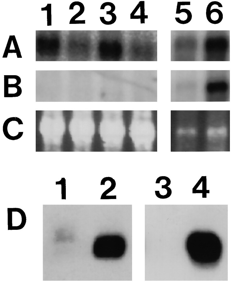
Fig. 2.
Analysis of the expression of PMP22 and P0. Northern blot analysis of PMP22-transgenic and wild-type mice (A–C). The same blot was hybridized first with a PMP22 cDNA probe (A) and subsequently with P0 cDNA probe (B). Ethidium bromide-stained agarose gel (C; 18S RNA) is shown as quantitation control. RNA was isolated from heart (lanes 1–4) and from sciatic nerves (lanes 5 and 6) of 21-d-old PMP22-transgenic mice (lanes 1, 3, and 5) and wild-type (lanes 2, 4, and 6) siblings (note that the exposure times of the different blots and probes were not identical). Western blot analysis of PMP22-transgenic and wild-type mice (D). Crude sciatic nerve homogenates (20 μg of protein) of PMP22-transgenic mice (lanes 1and 3; TgN248) and wild-type littermates (lanes 2 and 4) were separated on 12.5% SDS-PAGE and blotted to nitrocellulose membranes. Proteins were probed with antibodies specific for PMP22 (lanes 1 and2) or P0 (lanes 3 and4).

