
Fig. 4.
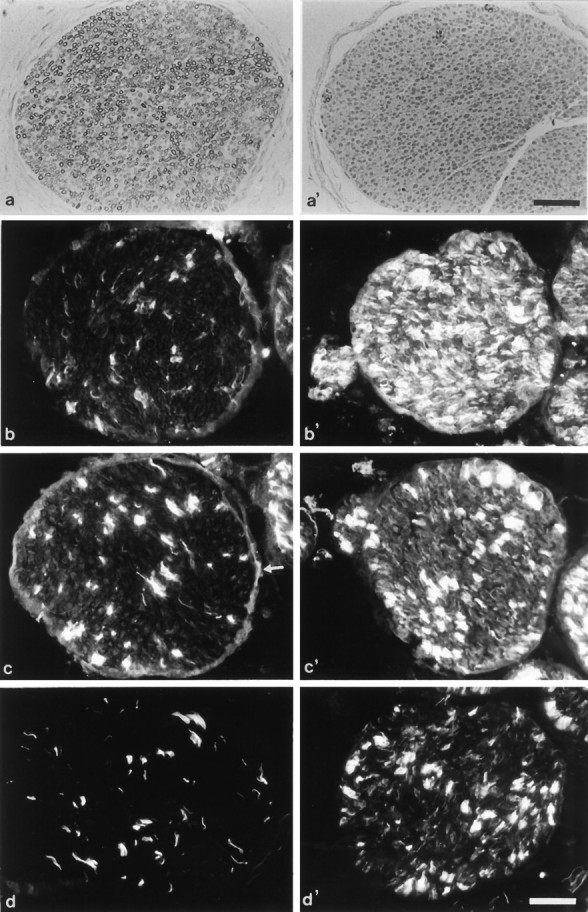
Assessment of myelination in semithin sections and immunohistological analysis. Cross-sections (2 μm) of 21-d-old wild-type (a) and PMP22-transgenic mice (a′) were stained with toluidine blue. Note the lack of detectable myelin (a′). Immunohistological localization of the cell surface molecules LNGFR (b,b′), N-CAM (c, c′), and L1 (d, d′) in femoral quadriceps nerves of adult wild-type (b–d) and mutant mice (b′–d′). Although LNGFR is prominently upregulated in mutant mice (b, b′), N-CAM is moderately (c, c′) and L1 weakly increased (d, d′). Note that the nonmyelinating axon-Schwann cell units also are labeled stronger for N-CAM and L1 in the mutants (c′, d′) than in the wild-type mice (c, d). Arrow inc marks the weakly N-CAM-immunoreactive perineurium of a wild-type nerve. Scale bars, 50 μm.