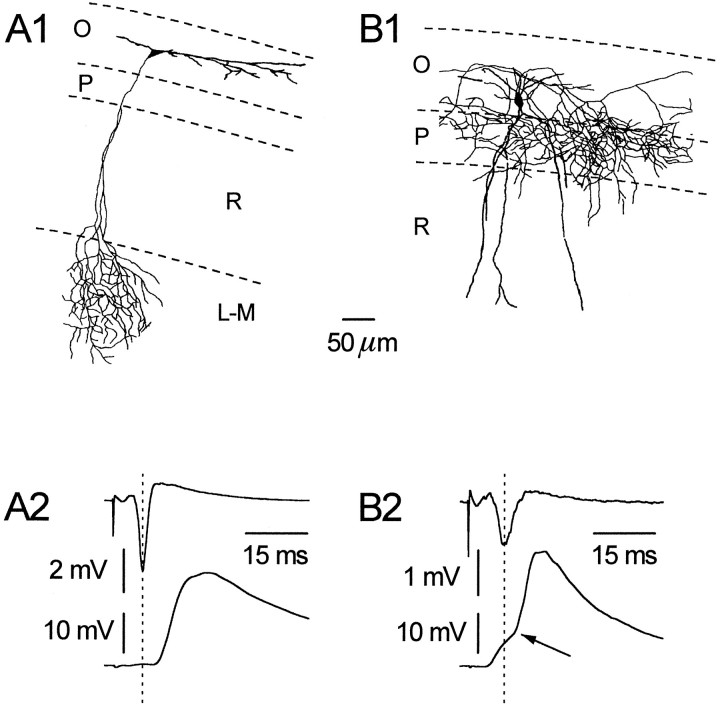
Fig. 1.
Horizontal and vertical OAIs can be differentiated both morphologically and electrophysiologically. A typical horizontal OAI is shown (A1); note its dendritic tree confined to st. oriens (O) and its axonal projection to st. lacunosum/moleculare (L-M). In A2, the result from a representative double-recording experiment is shown. After stimulation of st. radiatum (R) afferents, note that the peak of the fPS (upper trace) precedes the onset of the EPSP (lower trace) (+3.2 msec).B1 shows a vertical interneuron; note the dendritic arborization in both st. oriens and radiatum and the axonal plexus largely restricted to st. pyramidale (P).B2 shows a biphasic EPSP recorded from a vertical cell after afferent stimulation; note the early component preceding the fPS, whereas a second component follows it (−3.9 and +2.8 msec, respectively).

