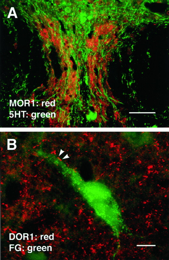
Fig. 11.

A, Section of nucleus raphe dorsalis stained for both 5HT and MOR1. As in nucleus raphe medianis (Fig. 9), serotonergic cells in NRD seldom if ever expressed MOR1-ir; however, MOR1-ir processes were found adjacent to 5HTcells and seemed to outline the region in which 5HT-ir cells occurred.B, DOR1-ir varicosities frequently apposed PAG cells retrogradely labeled from the RVM. In this image, FG was pseudocolored green. Scale bars: A, 50 μm; B, 10 μm.
