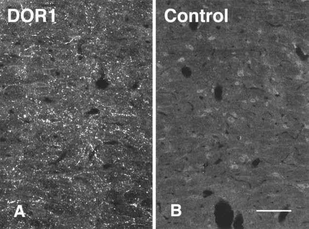
Fig. 5.

Images of adjacent sections through NRM stained with either the DOR1 antiserum (A) or the DOR1 antiserum to which was added 10 μg/ml of the peptide against which the antiserum was raised (B). Specific labeling was reduced or abolished in the absorption control. Scale bar, 50 μm.
