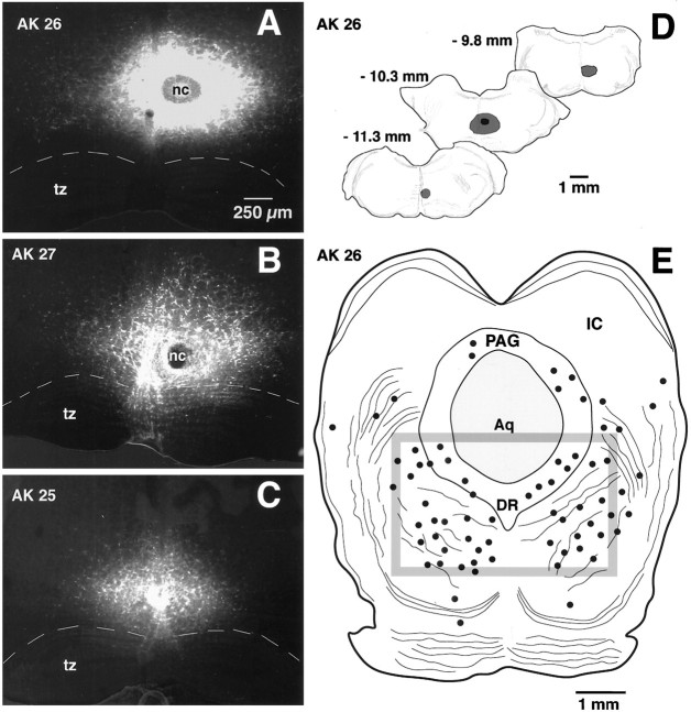
Fig. 7.
FG injection sites in the RVM and the resulting retrograde labeling in the caudal midbrain. A–C, Conventional images of FG injection sites in the RVM (largely NRM) for each of the three animals from which quantitative data were obtained (AK 26, AK 27, and AK 28). Each image is a montage of four to six individual micrographs. tz, Trapezoid body; nc, necrotic core. Dashed line represents the dorsal border of the trapezoid body.C, Camera lucida drawings of the rostrocaudal extent of the largest injection (AK 26). Numbersapproximate the level of the corresponding figures in the Paxinos and Watson atlas of rat brain. E, Retrograde labeling of neurons within a single section of the caudal midbrain of rat AK 26. Solid dots represent FG-labeled cells.Box represents the boundary of the region in which quantitative studies were performed. PAG, Periaqueductal gray; IC, inferior colliculus; Aq, aqueduct;DR, dorsal raphe.

